PET and SPECT imaging techniques are both detecting radioactive substances to visualize biological processes in the body. Although this sounds like they are similar, that is not the case. In this blog, we compare SPECT vs PET imaging. You will learn about sensitivity, resolution, and radionuclides. For drug developers, these factors play a crucial role in visualizing biodistribution and the uptake of drugs in the body. As an imaging CRO, TRACER is specialized in gathering first in-human data by labeling compounds for PET and SPECT. We compare PET vs SPECT imaging methods for each newly developed drug we visualize in vivo.
What is the basic mechanism of nuclear imaging?
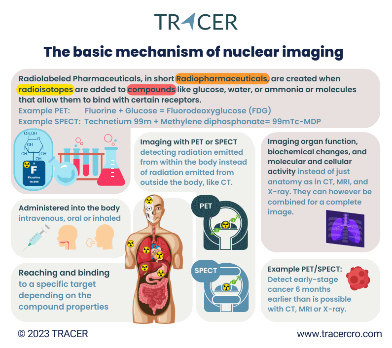
Nuclear molecular imaging uses radionuclides coupled to a targeting agent resulting in an imaging agent to visualize the biological process of interest in the body. In many diseases, specific biochemical processes or receptors are upregulated and can be used as a marker for disease activity. The disease activity can be visualized by selecting a ligand that is specifically metabolized by this biochemical process or a ligand that targets the receptors expressed on diseased cells. By labeling this ligand with a radionuclide (resulting in a radiotracer) it can be traced in the body with radionuclide imaging.
SPECT imaging vs PET in drug development: tracer – agent – ligand
The most widely used nuclear imaging techniques are Positron Emission Tomography (PET) and Single-Photon Emission Computed Tomography (SPECT). Making the comparison is all about choosing the combination of radionuclide, ligand, and imaging output.
SPECT vs PET, what do they have in common?
Let’s first look at the similarities between SPECT and PET. What are the common factors that you need to know when comparing SPECT vs PET scan? First of all, both are imaging techniques that detect radiotracers. Both also use tomographic imaging to generate a 3D image.
Differences between SPECT vs PET
Now that we’ve covered the similarities of PET SPECT, it is time to discuss the differences between SPECT versus PET. When selecting the imaging method for your clinical trial, it is important to understand the difference between SPECT vs PET. So, what are the strengths and weaknesses of PET SPECT? The difference between PET and SPECT consists of multiple factors, including technology, exposure to radiation, spatial resolution, temporal resolution, and binding capability.
PET SPECT difference in mechanism
To understand the differences, it is important to first understand the basic technology of PET and SPECT. We’ll cover this in the next two paragraphs.
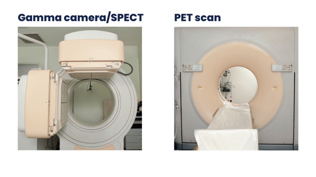
Comparing gamma camera vs PET scan, it is worth mentioning that PET has a more sophisticated detection method and algorithms to generate an image from the captured light of the radioactive tracer.
How does a PET scan work?
PET (Positron Emission Tomography) is a nuclear imaging technique that provides detailed functional and metabolic information about tissues and organs. For PET, positron emitting radioisotopes are used. Upon radioactive decay a positron is emitted. When a positron meets an electron in matter (for example the human body), annihilation takes place, and two gamma rays are emitted in opposite directions. These gamma rays are detected by a ring of detectors in the PET scanner that is surrounding the patient. The detected gamma rays can be reconstructed into 3D images. The 3D images visualize the distribution of the labeled substance in the body.
How does a SPECT scan work?
Single Photon Emission Computed Tomography (SPECT) is a nuclear medicine imaging technique that utilizes gamma ray-emitting radioactive tracers to visualize the functional activities within organs and tissues. SPECT usually consists of two crystal-based detectors that detect gamma rays and are placed opposite each other. Each detector generates a 2D image of the radioactivity distribution. By acquiring multiple 2D images at various angles, a 3D image can be computed.
PET vs SPECT resolution
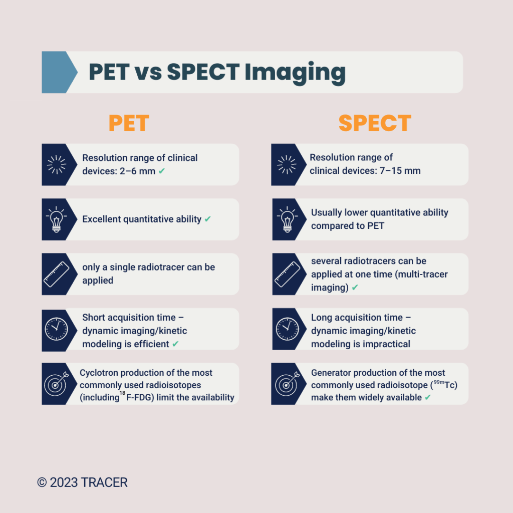
How does SPECT versus PET imaging compare in terms of spatial resolution? The resolution of PET images is higher compared to SPECT. At the same time, PET also has a higher sensitivity for the signals due to the different detection method used in PET scan vs SPECT scan. This is very useful in pharmaceutical R&D because low quantities of radioactivity can be made visible. Because for Phase 0 only a microdose of a drug is used, a PET scan can still generate a clear image.
Translating preclinical SPECT to clinical SPECT PET
There are tracers that can be used in preclinical SPECT or PET scans but are not intended for in-human use. In clinical studies, patient characteristics need to be considered when choosing a tracer and dosage.
Contact us as early as possible, even before the preclinical phase of drug development. By doing so, our scientists can help choose the correct radionuclide and labeling strategy and compare PET vs SPECT for your drug. This allows you to have consistent data from both your preclinical and clinical phases of drug development.
Discuss this with our scientist
PET and SPECT in drug development
- PET and SPECT are valuable tools in drug development and can be used for clinical research.
- With PET/SPECT, you can visualize PK/BD data of your drug compound. As PET offers more accurate quantification, this method helps you predict dosing and side effects in Phase 1 clinical trials.
- Personalized medicine and patient stratification: PET and SPECT scans can assist in identifying patients who are most likely to respond to a particular treatment.
- Target identification and validation with PET/SPECT by quantifying the presence and distribution of specific molecular markers.
- Evaluation of treatment efficacy: PET/SPECT imaging plays a role in evaluating the efficacy of drug candidates.
At TRACER, we have multiple PET imaging possibilities for the development of new drugs. Contact us for more information if you want to use PET imaging to generate in-human data in your drug development process.
Contact us
PET – SPECT advantages and disadvantages
- SPECT scanners and SPECT radioisotopes are more readily available.
- SPECT is less expensive compared to PET.
- PET offers a higher spatial resolution than SPECT.
- PET has a higher sensitivity, meaning smaller amounts can be detected than with SPECT.
- In general, SPECT radioisotopes can be measured from a couple of hours up to days, for PET this a couple of minutes to a couple of hours. However, there are exceptions, please read the explanation below.
- With PET more accurate quantification is possible, meaning you can get more accurate data on the accumulated drug doses. This can also be done with SPECT but less accurately.
PET vs SPECT radiation example
A PET scan with a zirconium-labeled antibody can give a higher radiation dose than a standard bone scan with SPECT. The dose depends on the amount of activity administered, the energy of the gamma photons and the time the radionuclide is present in the body. So, it is not standard that PET always gives less radiation dose than SPECT.
Choosing the right tracer to reach the target in the body
Different radiotracers in SPECT and PET can visualize different compounds and organs. The following two examples show the necessity of choosing the correct label for measuring certain organs.

Brain PET and brain SPECT imaging
For both brain PET and brain SPECT imaging there is a specific factor that should be considered when choosing a tracer. Not all tracers are able to cross the Blood-Brain-Barrier (BBB). Depending if the tracer is more hydrophilic or lipophilic and depending on the transport mechanism, the tracer may be able to pass through the BBB or not. What is the difference between PET and SPECT brain scan? The same differences between PET and SPECT do still apply. Are you considering options for SPECT vs PET brain imaging for a new drug? Please contact us so our scientist can provide you with advice.

Cardiac SPECT vs PET
Cardiac PET images and SPECT images are useful for cardiologists. By making a PET image or SPECT of the heart, different blood flows can be visualized and analyzed. What is the outcome of comparing SPECT vs PET cardiac imaging?
- The longer half-life of SPECT radiotracers makes it more favorable in cardiology.
- The image quality is the main reason to choose cardiac PET imaging.
- Cardiac stress testing can be performed with PET and SPECT scans.
- SPECT myocardial perfusion imaging is used to evaluate the damage caused by myocardial infarction (for example reduced blood flow). This SPECT test of the heart is commonly used in cardiology.
- Cardiac PET/SPECT scans can be done gated. Read at the end of this article more about SPECT heart imaging in the question about gated SPECT test/PET stress test.
Commonly used tracers for PET vs SPECT
In PET scans different tracers can be used. Halogen isotopes such as fluorine-18, iodine-131, and carbon-11 can be used in PET scans when labeling compounds. Radiometals are copper-64, gallium-68, and zirconium-89.
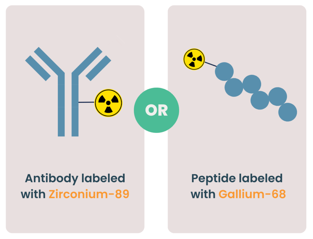
It is important that you match the in vivo half-life of your compound with the physical half-life of the radioisotope. For example, zirconium-89 with a half-life of approximately 3 days can be used to label antibodies with a long circulation in plasma for PET scans. The short half-life of gallium-68 (68 minutes) can be used to label peptides, which have a fast clearance from the body, for PET imaging commonly used SPECT tracers are for example, technetium-99m, Iodine-123, and indium-111.
Do you want to know which isotope is the best for labeling your compound? Contact TRACER for more information.
PET and SPECT radiopharmaceuticals in relation to the application
The choice of the radiotracer is depended on the binding factors of your target. In the case of using PET and SPECT in clinical research, it depends on the binding factors of both target and the investigated compound. The example below of gallium-68 supports this.
Gallium 68 in combination with different peptides
Gallium 68 is a radioisotope of gallium. Octreotide is a specific peptide with binding to somatostatin receptors, found in high levels in neuroendocrine tumors. Several octreotide analogs (DOTATOC, DOTATATE, DOTANOC) are labeled with gallium-68 for PET imaging. Each analog binds with a different affinity to subfamilies of the somatostatin receptor. As a result of that, each analog has its own specific biodistribution in patients with neuroendocrine tumors.
Understanding the choice of tracers for different targets
The below list gives a good understanding of how different tracers can interact with the target.
- For a Rubidium cardiac PET scan Rubidium-82 is used. Rubidium-82 is made from Strontium-82 with a generator. Rubidium has a high uptake in the heart and is therefore used in a pet scan for heart studies.
- Technetium-99m is used in SPECT with a large variety of Tc-99m-labeled radiopharmaceuticals for various diseases in many different organs. The broad possibilities of application and high availability make Tc-99m a widely utilized radiotracer.
- HMPAO is short for Hexamethylpropyleneamine Oxine, a radiopharmaceutical agent used for SPECT, particularly in brain perfusion imaging. It is basically the same as Technetium (99mTc) exametazime with only some slight differences in chemical structure.
- Thallium 201, a radiopharmaceutical agent, is used in Thallium SPECT to study coronary artery disease (Thallium stress test) and parathyroid hyperactivity. There are studies using Thallium-201 in oncology and olfacto-scintigraphy.
- Tc-99m-TRODAT-1 binds to dopamine transporters and is imaged with a SPECT scan.
- Ga-68-PSMA is used for a PET scan to measure PSMA-expression in prostate cancer patients.
- Lutetium-177-PSMA is used for radionuclide therapy in prostate cancer patients. The accumulation of the radiopharmaceutical can be visualized and quantified by SPECT for therapy response monitoring and dosimetry.
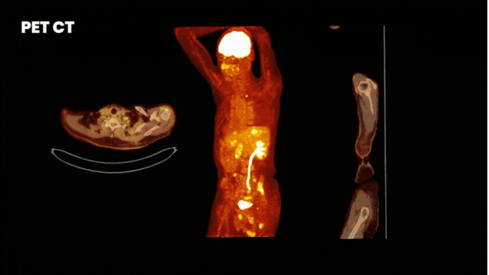
SPECT CT and PET CT
Both imaging methods can be combined with CT (Computed Tomography). A complete image of anatomy and physiology can be made in one scan with SPECT CT and PET CT. Does combining SPECT or PET with CT change the method itself? No, for instance with a SPECT/CT scan the radioactive tracer is still used for the gamma camera, and CT is using its own X-ray radiation to generate data on the anatomy. This way both the target of the PET or SPECT scan and its exact anatomical location can be determined. This is very useful for drug development R&D for measuring the presence and accumulation of a compound in the body.
SPECT in comparison to CT and MRI
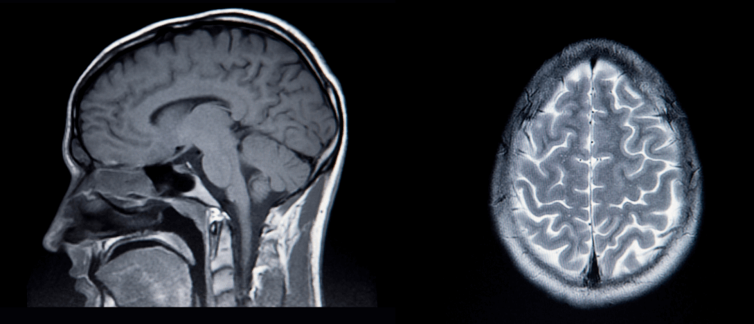 MRI scan
 CT scan
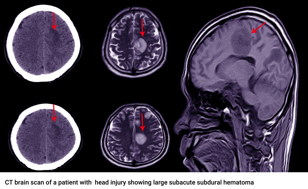
The advantage of using nuclear molecular imaging is that it provides information that cannot easily be obtained with standard radiology imaging techniques (e.g. CT or MRI), as these techniques usually measure the size and morphology of the lesion and not the actual biological process or target.
Example: using PET or SPECT in cancer treatment monitoring
A clear example can be found in cancer treatment monitoring. The treatment might not initially result in shrinkage of the tumor (i.e. imaged by CT or MRI) but can result in a strong reduction in biochemical processes and an indirect measure of effect. This effect can be measured by nuclear and optical molecular imaging. By only measuring tumor volume through CT or MRI, it can then be incorrectly concluded that the therapy is not having an effect. However, by using PET or SPECT imaging a treatment effect would be observed.
It is possible to perform functional imaging with MRI by using MRI contrast agents. Due to the sensitivity of PET and SPECT, these imaging modalities are favorable for molecular imaging. The big advantage of MRI is that it does not require the use of ionizing radiation.
FAQ
In short, we answer the most important questions covered in this article. In case you have a question about SPECT, PET, tracers, or any other subject related to drug development, don’t hesitate to contact us.
What is PET and SPECT?
PET (Positron Emission Tomography) and SPECT (Single Photon Emission Computed Tomography) are both imaging techniques that use radioactive tracers. For PET positron-emitting tracers are used. Gamma rays are generated when the tracers interact with electrons in the body. For SPECT gamma-emitting radiotracers emit gamma rays directly and can be detected with a gamma camera.
What is gated SPECT?
What is gating in imaging? And what does gated mean in gated SPECT? Compared to normal SPECT, which just takes static images, gated means the moment images are taken is controlled. A good example is gated SPECT myocardial perfusion imaging, a method for SPECT tests heart perfusion and function. The images are taken at specific moments, on contraction or relaxation of the heart. Based on the ElectroCardioGram (ECG) the ‘gates’ are set where an image should be made. Good to know, gated does not only exist with SPECT. Also, gated cardiac imaging with PET is possible where it is commonly used for respiratory gating (to reduce artifacts by the movement of the body by breathing). Gated imaging is not limited by a specific type of tracer, many different tracers can be used.
Why use SPECT over PET?
The high availability and large number of different radiotracers are one of the main reasons to use SPECT over PET. Also, the costs are generally lower for SPECT than for PET. Contact TRACER for an indication of the costs of using SPECT or PET for clinical research.
Why is PET scan better than SPECT?
Why is PET better than SPECT? PET:
• offers higher spatial resolution;
• has a higher sensitivity and specificity, meaning smaller amounts can be detected;
• quantification is more accurate.
Which is better PET or SPECT?
Comparing PET and SPECT imaging and choosing the right method for drug research is highly dependent on your research question. When a high resolution is needed, PET might be the logical choice, but the resolution is not the only factor to consider. For instance, when choosing between a PET scan and SPECT scan to get in-vivo data on a new drug, the radionuclide needs to be able to conjugate with the compound. SPECT can be especially useful in radionuclide therapy. Some beta-emitters used for therapy such as Lutetium-177 (Lu-177) and Iodine-131 (I-131) also emit a gamma photon. This gamma photon can be used for SPECT.
When developing a radiopharmaceutical for therapy, SPECT imaging can be used for patient selection, therapy response monitoring, and dosimetry, without the need for a separate diagnostic imaging agent.
As a drug developer or biotech company, you can discuss with TRACER’s scientists what radiotracers are most suitable for your compound and research question.