Raman imaging is a powerful analytics method in drug development. Raman spectroscopy and Raman microscopy can be used in both drug discovery and clinical drug development. With the Raman scattering effect, the changes in the wavelength of light and energy can be analyzed and used to identify molecules in solids, liquids, and even gasses. The Raman effect occurs when light hits a molecule that in reaction rotates and vibrates. A Raman analysis is done with Raman spectroscopy instrumentation, where the chosen laser Raman spectrophotometer depends on the molecule to analyze.
Possibilities of a Raman instrument in clinical trials
How can Raman spectroscopy be used in drug discovery, preclinical drug development, and clinical trials? Almost all molecules, except for fluorescent molecules, can be detected and identified with Raman scattering. This allows for a thorough understanding of drug-related effects and toxins. Regular Raman spectroscopy does not require labeling and little to no sample preparation. For Surface Enhanced Raman Spectroscopy (SERS), nanoparticles can be coupled with compounds such as antibodies for imaging of certain targets. DNA can also be analyzed, even circulating tumor DNA [1].
Can Raman be used for my drug development?
As an expert in molecular imaging in clinical trials, we at TRACER can help you set up and conduct any clinical trial with Raman spectroscopy. Especially for dermatology clinical trials, this is often a very suitable option. The uptake of the topical-delivered drug can be analyzed with Raman in a non-invasive procedure. This is more convenient for the patient and regulators also view this positively. No labeling of the compound is required. Raman is suitable for many different molecules. Depending on the type of Raman, this method is highly sensitive. SERS can be used for concentrations too low for regular Raman imaging.
Utilize Raman in your clinical trial
A Raman instrument offers molecular pharmaceutical analysis on disease pathology, also allowing for the identification of possible disease endotypes.
Benefits of Raman in clinical trials
- Use Raman scattering to evaluate the presence of biomarkers and to understand the cellular structures.
- Raman can be used to detect molecules in solids, liquids, and even gases.
- Discover possible changes in your drug formulation after compound uptake.
- Raman spectroscopy can detect known molecules and identify the unknown ones with spectra stripping combined with libraries.
- Raman instruments are very suitable for analyzing topically delivered drugs or measuring uptake in a sample.
- Raman microscopy is a non-destructive, and non-invasive imaging method.
- The limited penetration depth of Raman microscopy, 1–6 µm, should be taken into consideration.
- Raman can be used in combination with other imaging methods, but interference – such as from fluorescence – should be considered in the imaging strategy.
Design your imaging strategy with TRACER or ask questions about Raman for your clinical trial.
Raman spectroscopy in exploratory clinical trials
Raman analysis can be very useful in drug development, especially in the exploratory phase of clinical trials. Exploratory clinical trials are the first in-patient trials, that can be conducted before Phase 1, replacing part of the preclinical development. Drug developers can learn more about their compound and disease characteristics in-patient by using the possibilities of exploratory trials, also known as Phase 0. Raman is a valuable technology worth considering before starting your clinical trials. Allowing you to obtain more information, from the same clinical trial.
Smarter clinical trial design
Exploratory trials with imaging provide a better understanding of disease characteristics. This can enhance Phase 1 in-patient and Phase 2 trial design. Biological imaging of chemical bonds by stimulated Raman scattering microscopy can provide knowledge on the behavior and effects of the compound in-patient. Depending on your exploratory clinical trial design, the obtained data can be used for lead candidate selection, go/no-go decision-making, and subsequent trial design. As an alternative or addition to Raman Imaging, modalities such as PET/SPECT and fluorescence can be considered.
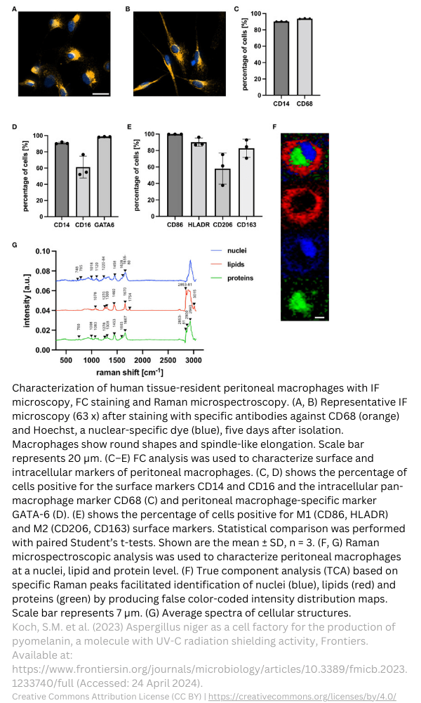
Raman spectroscopy basics
Raman scattering spectroscopy is basically the same for all Raman variations. There is a photon-molecule interaction, where the scattering of light can be used to analyze chemical composition and molecular structure. The rest of this article will explain the basics of Raman scattering and Raman spectroscopy.
Raman spectroscopy explained
What is the principle of Raman spectroscopy? Raman makes use of a laser light that is pointed at a sample. Depending on the shape and energy of a molecule, each molecule has a different way of vibrating. The molecule can then absorb the light, scatter it, or transmit it. The Raman effect is scattering of light. The light can scatter in two forms which we explain in the following paragraphs.
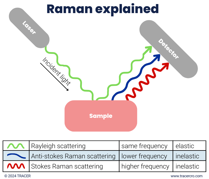
Raman elastic scattering
The first one is elastic scattering (in Rayleigh green) where there is no net change in photon input and photon output. When a light is shined on a molecule it goes into a virtual state and falls back to the ground state. The wavelength of the photon, input and output, is equal. This is called elastic scattering.
Raman inelastic scattering
The second way is inelastic scattering or Raman scattering (in blue and red). Inelastic scattering means there is a difference in photon input and output. Raman scattering forms a unique fingerprint of the molecule. With Raman scattering, the molecule does not fall back to the initial state but falls into a lower or higher vibrational state. The wavelength of the released photon after passing the molecule can be lower or higher than its initial wavelength.
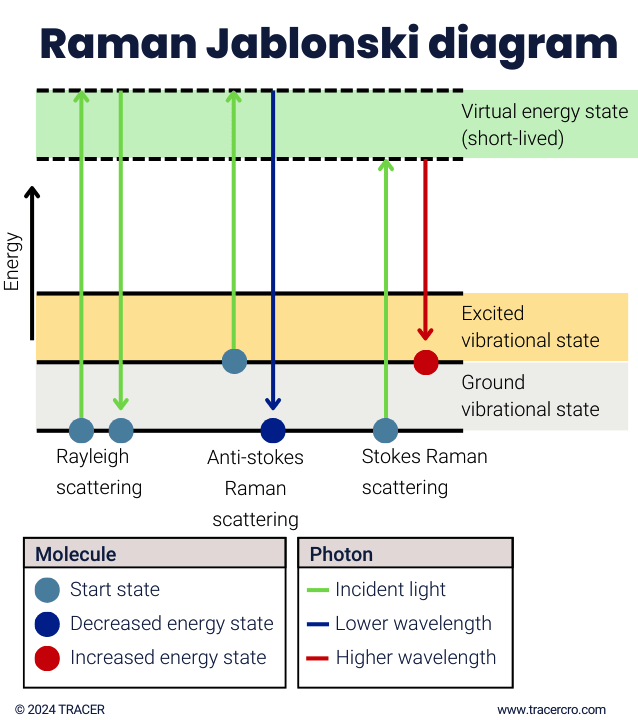
Raman scattering: stokes or anti-stokes
Raman spectroscopy utilizes the Raman stokes scattering. There are two ways of Raman scattering. They are dependent on the interaction of the photon with the molecule and vice versa.
- A large difference between input and output photons shows that the molecule has a high-frequency vibration of light atoms with strong bonds.
- When a small Raman change is observed, this is often an indication of heavy atoms with weak bonds.
|
Released photon |
Molecule |
||||
|
Type |
Wavelength |
Energy |
Start state |
End state |
Vibrational state |
|
Elastic scattering |
Same |
Same |
Ground |
Ground |
Same |
|
Stokes Raman |
Higher |
Decreased |
Ground |
Excited |
Higher |
|
Anti-stokes Raman |
Shorter |
Increased |
Excited |
Ground |
Lower |
Raman stokes scattering
The first form of Raman scattering is stokes Raman scattering. The molecule goes from ground state to virtual state and then to excited state. In other words, from a low to a high vibrational energy level. The released photon has less energy and a longer wavelength than that of the initial photon.
Raman anti-stokes scattering
The other way of Raman scattering is anti-stokes Raman, where the molecule goes from an excited state into virtual state and then falls back to a ground state. The molecule goes from a high to a low vibrational energy level. The output photon has gained energy, resulting in a shorter wavelength.
Raman analysis
The Raman scattering output can be analyzed in multiple ways. The characteristic frequencies help to determine the material composition. Changes in peak position indicate stress and or strain state. The intensity of the peak indicates the amount of material, a higher peak indicates a higher concentration. The width of the peak can indicate in some materials the quality of that material. Finally, the polarization can be parallel or perpendicular indicating the orientation.
Raman spectrometer
A Raman spectrometer is used in Raman spectroscopy. The setup consists of a laser, spectrometer, and sample. The laser shines on a beam splitter and is projected through an optic onto the sample. The beam is then reflected through another optic and a filter before entering the spectrometer. The Raman spectrometer consists of an entrance slit, lens, and grating. The grating splits the light into the wavelengths and sends it to a Charge Coupled Device (CCD) detector. This device creates the Raman lines, or in other words, the Raman spectrum.
Raman spectrometer often only uses Raman stokes
Commonly only the Raman stokes scattering is used for analysis because anti-stokes Raman is weaker than stokes. The much stronger Raleigh wavelengths are filtered out completely to prevent interference of the stokes Raman signal.
Raman spectroscopy: good to know
In Raman spectroscopy, a mode refers to the vibrational motion of atoms. Molecules can have multiple modes. Modes can include bending, stretching, or twisting. The frequencies of vibrational modes give information on the chemical composition, molecular structure, and physical properties. Infrared (IR) spectroscopy and Raman spectroscopy can be used to gain information on molecular structure, composition, and interactions between molecules. IR and Raman can have overlapping measurements, but some measurements can only be done with one or the other.
Raman spectroscopy instrumentation
There are different Raman instruments. The handheld Raman spectrometer is used to quickly identify compounds, like for goods control. Fourier Transform FT Raman or FT Raman spectroscopy is a device to automate sample analysis. For a more detailed image or in-depth Raman spectra analysis, a Raman microscope can be used. Examples are the inVia Raman microscope and the DXR3 Raman microscope. Note that not every Raman measurement tool is the same. The Raman shift measured is relative to the laser used in the Raman instrumentation. Since the shift is relative, the Raman spectra analysis will have the Rayleigh in the middle, the stokes right, and the anti-stokes on the left.
Types of Raman imaging
Over the years, many variations of Raman imaging have emerged, solving problems with spatial resolution, sensitivity, and specificity, and adding imaging capabilities. Below we list common Raman variations.
- Spontaneous Raman Spectroscopy (traditional Raman)
- Raman microscopy (µ-Raman)
- Stimulated Raman Scattering (SRS)
- Surface Enhanced Raman Spectroscopy (SERS)
- Resonance Raman Spectroscopy (RRS)
- Surface-Enhanced Resonance Raman Spectroscopy (SERRS)
- Spatially Offset Raman Spectroscopy (SORS)
- Coherent Anti-Stokes Raman Spectroscopy (CARS)
- Tip-Enhanced Raman Spectroscopy (TERS)
- Time-Resolved Resonance Raman Spectroscopy (TR3S)
Polarized Raman spectroscopy
There is one major difference worth mentioning in laser Raman spectroscopy. Polarized Raman is different from the discussed Raman technology above. It gives additional information on the vibration and rotation levels. By using a polarized laser instead of an unpolarized laser the anisotropic properties can be measured. This is done by measuring the intensity and direction of the scattered light.
Surface Enhanced Raman Spectroscopy
Surface Enhanced Raman Spectroscopy, or in short SERS Raman enhances the signal and provides a more sensitive reading than normal Raman spectroscopy. This technique can be used in drug discovery and to identify cancer cells. Due to high sensitivity and specificity, specific biomolecules can be identified and analyzed. SERS Surface Enhanced Raman spectroscopy uses different instrumentation. SERS requires coupling your compound with nanoparticles. For in vivo Raman spectroscopy, these particles need to be biocompatible and not interfere with the compound’s behavior. Surface Enhanced Raman Scattering is not always needed. We can discuss your project and help you determine the needed setup. Contact us to schedule a meeting with our scientist.
Frequently asked questions
Below we answer some of the frequently asked questions about Raman spectroscopy. If you have a different question, or if you would like to learn more about the use of Raman microscopy or spectroscopy in clinical trials, please contact us.
What Raman spectroscopy is used for?
Raman is used to identify molecules. It can identify unknown substances when combined with software to compare the measured substance with a library. There are many Raman spectroscopy applications. At TRACER CRO we focus on the clinical use of Raman, but application in laboratories and the industry is common.
What are the possibilities of Raman spectroscopy of biological tissues?
In clinical trials, the Raman spectroscopy pharmaceutical analysis go beyond measuring drug uptake. Changes in cells induced by the compound can be measured. With Raman, it is possible to measure for example proteins, lipids, and nucleic acids within a cell. Furthermore, it is possible to gain insight into diseased tissue.
What is Raman microscopy used for?
Because of the molecular level of analysis, Raman is used in science, chemistry, biology, and medicine development. In drug discovery, Raman imaging can be used to develop a formula for compounds. In drug development, the interaction of the drug and the target can be measured by analyzing the Raman effect. At TRACER we can do this for your drug or molecule. By using Raman microscopy, a chemical image of the sample in cells and tissues can be made. A big difference between Raman spectroscopy and Raman microscopy is the size of the measurements. As you might expect, with Raman microscopy individual cells and sub-cellular structures can be analyzed.
What are the advantages of Raman spectroscopy?
Raman spectroscopy instrumentation can analyze substances in transparent, sealed containers as well as in water. It is a non-destructive technique that often requires no sample preparation. Polymorphs, the same atoms in different crystallization forms, can be measured. When considering Raman in clinical trials, it is worth mentioning that labeling your compound is not needed.
What is the difference between IR and Raman spectroscopy?
Although Raman and IR fall both under vibrational spectroscopy, they differ from each other. IR works with the absorption of light, while Raman works with light scattering. Raman and IR are complementary techniques. For instance, aqueous substances are difficult to analyze with IR due to infrared absorption. Since water itself has a low Raman scattering, substances in water can be analyzed with Raman. IR can be used to analyze fluorescent substances that provide too much background noise for Raman.
What is the difference between Raman and FTIR?
The previous paragraph has the difference FTIR Raman explained in short. Fourier Transform Infrared Spectroscopy (FTIR), is similar to IR, only a broad range of wavelengths can be analyzed simultaneously. This makes FTIR faster and more sensitive than IR. FTIR and Raman spectroscopy are, like IR and Raman, complementary to each other.
Abbreviations
|
SERS |
Surface Enhanced Raman Spectroscopy |
|
CCD |
Charge Coupled Device |
|
IR |
InfraRed |
|
FT |
Fourier Transform |
|
SRS |
Stimulated Raman Scattering |
|
RRS |
Resonance Raman Spectroscopy |
|
SERRS |
Surface-Enhanced Resonance Raman Spectroscopy |
|
SORS |
Spatially Offset Raman Spectroscopy |
|
CARS |
Coherent Anti-Stokes Raman Spectroscopy |
|
TERS |
Tip-Enhanced Raman Spectroscopy |
|
TR3S |
Time-Resolved Resonance Raman Spectroscopy |
|
CRO |
Contract Research Organisation |
|
FTIR |
Fourier Transform Infrared Spectroscopy |
References
Although this article has been composed with great care and attention, we cannot guarantee its accuracy. If you have any suggestions or additions to this article, please email info@tracercro.com.
No rights can be derived from this publication. This blog does not make claim or promote ownership to any intellectual property, study information, clinical images, or copyrighted terms wherein.